The Neuromuscular Junction
For many years the most intensively studied synapse was the neuromuscular junction (NMJ). This is a synapse between a motor neuron and a skeletal muscle fiber that is physically quite large and accessible, making it relatively easy to study (Figure 1). The person who is most closely associated with these studies is Bernard Katz. It is rare in science for someone to so completely dominate a field as important as synaptic physiology in the way that Katz did for two decades between 1950 and 1970. A large part of everything that we know about synaptic transmission was first discovered or suggested by Katz. He received the Nobel prize in 1970.
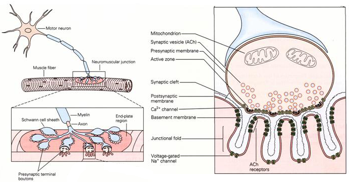
Figure 1 Structure of the neuromuscular junction.
The neurotransmitter at the neuromuscular junction is acetylcholine (ACh). At this synapse, ACh is an excitatory neurotransmitter, meaning that it depolarizes the cell membrane. The neuromuscular junction is made up of multiple synaptic boutons, making it an unusually large synapse (Figure 1). There are large, junctional folds in the postsynaptic membrane positioned opposite to the active zones in the presynaptic membrane. At the crest of each fold AChRs are clustered in a very high concentration (∼10,000 receptors per square micrometer). The active zone contains voltage-gated Ca2+ channels.
In his studies on the neuromuscular junction Katz made numerous important contributions including:
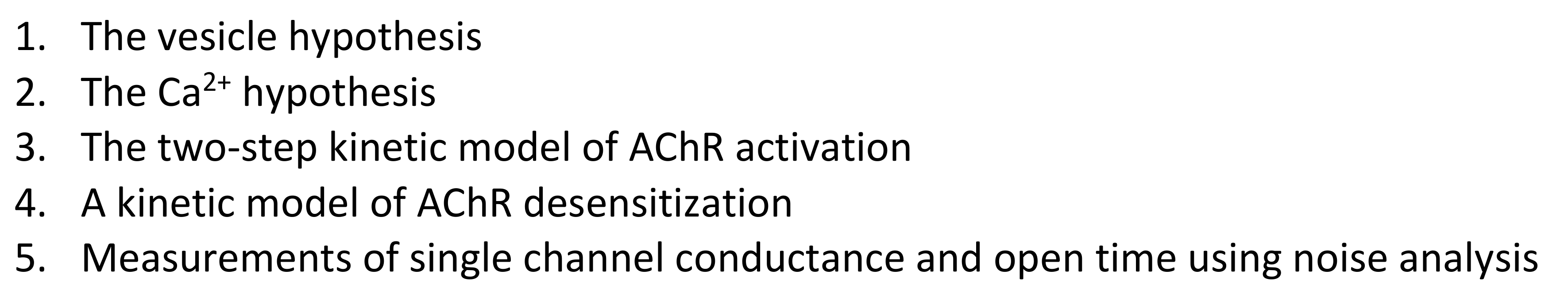
A typical experiment performed by Katz and his colleagues involved stimulating the axon of the presynaptic nerve and then recording the voltage change in the postsynaptic cell (Figure 2). One of his most important experiments involved nothing more than turning up the gain on the recording amplifier.
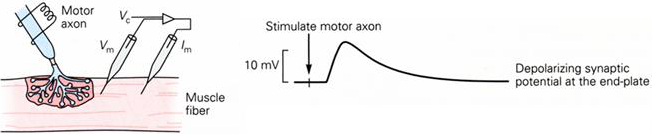
Figure 2 Experimental set-up for recording from the neuromuscular junction (left panel). Excitatory postsynaptic potential recorded following nerve stimulation (right panel).
Original recordings from an experiment by Katz are shown in Figure 3. In the bottom trace is a recording of an epsp and subsequent action potential in the muscle fiber. In the upper traces the amplifier gain is set much higher and the small blips in the traces are spontaneous epsps. They tend to be all much the same size and occur spontaneously and randomly without any electrical excitation.
At the same time as Katz was recording his miniature epsps other scientists were starting to use electron microscopes to study biological samples. When they looked at the neuromuscular junction they saw that the nerve terminal was filled with hundreds of small spherical structures (Figure 3).
Figure 3 Spontaneous miniature end-plate potentials (left panel). Electron micrograph of vesicles in the nerve terminal (right panel).
In 1955 Katz put these two observations together and surmised that the vesicles seen in the electron micrographs were filled with neurotransmitter and that the miniature epsps were due to the spontaneous fusion of these vesicles with the membrane. In his vesicle hypothesis, a normal epsp occurred when an action potential invaded the nerve terminal and caused a large number of vesicles to be released. He also anticipated that there would be a specialized docking site where the vesicles attached to the nerve terminal membrane prior to release.
The second major contribution Katz made was to begin to understand how the electrical signal in the nerve terminal induced the release of neurotransmitter. In 1965 he again performed a critical experiment that was both conceptually and technically quite simple. Katz removed all the extracellular Ca2+ ions from the external media. Under these conditions synaptic transmission completely fails. He then brought a pipette up close to the synapse and allowed a small amount of calcium to leak out of the pipette. Under these conditions synaptic transmission is partially restored. More external Ca2+ produced more neurotransmitter release (Figure 4).
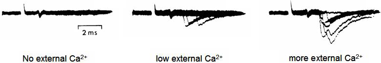
Figure 4 Effect of increasing external Ca2+ concentration on neurotransmitter release.
These two basic concepts, the vesicle hypothesis and the observation that calcium ions are the link between excitation and secretion are central to our understanding of synaptic transmission.
The Neuromuscular Junction is a Large Synapse
The NMJ is an unusual synapse in that synaptic transmission is 'fail-safe', it always works except under extreme conditions. This is because the synapse is so large. A large number of vesicles and, consequently, a large amount of neurotransmitter is released by a single action potential invading the presynaptic terminal and there are a very large number of receptors available to bind the neurotransmitter and then open. The resultant epsp is of the order of 70 mV in size (Figure 5).
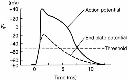
Figure 5 Excitatory postsynaptic potential (also known as end-plate potential) at the neuromuscular junction.
The epsc underlying the epsp at the neuromuscular junction is approximately three times larger than needed to cross the threshold for action potential generation. There is a non-linear relationship between epsc size and epsp size so the epsp does not look this large in Figure 5. This excess epsc size is known as the safety margin and provides a measure of security at neuromuscular synapses during stress or disease. A failure of synaptic transmission at the neuromuscular junction on the diaphragm muscle controlling the lung results in a cessation of breathing and rapid death. Failure at other neuromuscular junctions results in immobilization. Neuromuscular junction toxins are used by predators in the natural world to stun or kill prey e.g., the snake venom toxin α-bungarotoxin.