Basic Features of Synaptic Transmission
Synapses are the single most characteristic feature of neurons. Neurons in the mammalian central nervous system typically have from 100 to 100,000 synapses per cell. In a human brain there are a total of approximately 1014 synapses. From one perspective, neurons are simply the cellular scaffolding that allows the expression of synapses.
The synapse is a highly specialized cellular structure that facilitates rapid communication between two electrically excitable cells. The transfer of electrical excitation is a one-way process; from the presynaptic nerve terminal to the postsynaptic cell. Although the transmission of electrical signals is unidirectional, chemical signals that maintain the synaptic contact and modulate its strength can pass back and forth between the pre- and post-synaptic cells.
Most synapses are chemical synapses, which means that the presynaptic cell releases a signaling molecule known as a neurotransmitter. This neurotransmitter molecule then diffuses towards the second cell where it binds to a neurotransmitter receptor. Binding of the neurotransmitter then triggers a response in the second cell via activation of the neurotransmitter receptor.
The unique geometry of the synapse has two functions: it reduces diffusion times and increases the specificity of signaling. The time taken for molecules to diffuse through solutions is a common limitation for biological organisms. Much of the architecture of different organ systems can be explained by the necessity to reduce diffusion times. Diffusion times increase in proportion to the square of the distance over which the molecules diffuse. As a consequence, reducing the distance for diffusion has a dramatic effect on the time taken for diffusion. The synapse brings the membrane of the pre- and post-synaptic cells into very close apposition to minimize diffusion times. Typically, there is a gap of 10 to 40 nm between the pre- and post-synaptic membranes resulting in a diffusion time of significantly less than 1 millisecond. Bringing the presynaptic terminal so close to the postsynaptic cell also increases signaling specificity, so that at most fast synapses only the two cells making the synapse are in communication. This is very different to how other signaling molecules, such as hormones or cytokines, are used. Hormones for example are released into the blood stream and can affect a broad number of cells due to their diffuse distribution pattern.
Synaptic Structure
The synapse is an unusual subcellular structure in that it is made by a collaboration between two different cells that have to communicate with each other in order to successfully form the synapse. They each have to contribute several different structural components to the synapse to make it work. Typically, several signaling systems in addition to normal synaptic transmission are involved in the back and forth communication between the two cells in order to correctly assemble the synapse.
The locations of the main structural elements found at a typical synapse are shown in (Figure 1). The nerve terminal is filled with small spherical structures known as synaptic vesicles. These are membrane-bound spheres that are filled with neurotransmitter. The nerve terminal also has specializations on the surface facing the post-synaptic cell known as vesicle docking sites or active zones. These are the points where the vesicles bind and then fuse with the cell membrane to release neurotransmitter into the synaptic cleft. Also, in the presynaptic membrane are voltage-gated calcium channels. Located on the post-synaptic side are the neurotransmitter receptors and associated molecules that are collectively known as the postsynaptic density.
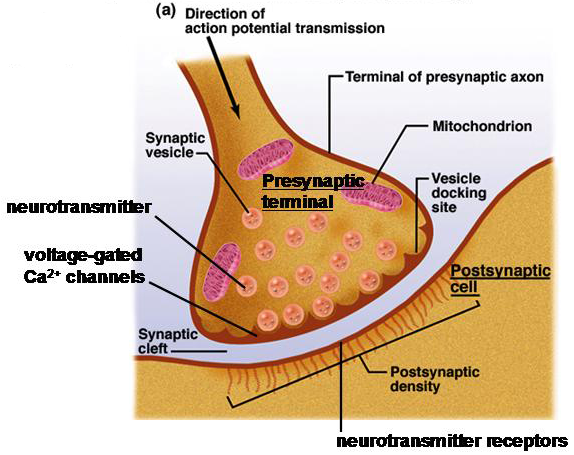
Figure 1 Structural features of a typical synapse.
Different Types of Synapses
There are many different types of synapses. The nature of different synapses is determined by the kind of the neurotransmitter that the pre-synaptic cell releases and the type of neurotransmitter receptor found in the membrane of the postsynaptic cell. It is usually most useful to categorize synapses on the basis of the kind of neurotransmitter receptor that it expresses.
Ligand-gated ion channels can be either excitatory or inhibitory. Excitatory receptors allow cations to pass (typically Na+ and K+, and sometimes Ca2+) through their integral ion channels. Opening of these channels depolarizes the cell membrane potential bringing the membrane potential closer to the threshold for action potential firing (Figure 2). This is known as an excitatory postsynaptic potential (epsp). The integral ion channel of inhibitory receptors is typically permeable to Cl- ions, the major extracellular anion. Activation of these inhibitory receptors moves the membrane potential away from the threshold potential. This is known as an inhibitory postsynaptic potential (ipsp).
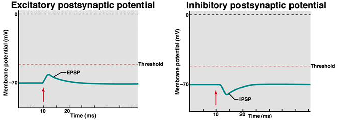
Figure 2 Effect of opening excitatory, cation channels or inhibitory, anion channels on membrane potential.