Regulation of Internal Ca2+ Ion Concentration
Intracellular Ca2+ ion concentrations are maintained at very low levels inside the cell and there is a very large concentration gradient of Ca2+ ions across the cell membrane (Table 1).
Table 1 Calcium ion concentrations inside and outside of cell
| Ion | Intracellular | Extracellular |
|---|---|---|
| Ca2+ | 100 nM | 1 mM |
There are a multitude of transport systems that contribute to the regulation of the internal Ca2+ ion concentration (Figure 1). The Ca-ATPase pump is found in the cell membrane and several organelle membranes. It actively pumps Ca2+ ions out of the cell or into intracellular organelles (mitochondria and endoplasmic reticulum), where the Ca2+ ions are sequestered. In addition, there is the Na-Ca exchanger that uses the energy of the Na+ ion gradient to transport Ca2+ ions out of the cell. Calcium binding proteins are another important part of the system. These proteins bind Ca2+ ions and act like buffers, rapidly reducing free Ca2+ ion concentrations following an influx of Ca2+ ions through open calcium channels. These buffers give the more slowly acting pump and transporters some time to remove the ions from the intracellular fluid. The buffers limit the duration and extent of the change in free Ca2+ ion concentration within the cell.
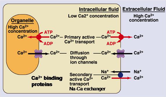
Figure 1 Summary of the various systems for handling Ca2+ ions within the cell.
Ca2+ Ions as Second Messengers
Perhaps oddly, given the effort that cells expend to keep internal Ca2+ ion concentrations low, Ca2+ ions have an important role as intracellular signaling molecules. Although an apparently unlikely candidate, given the simple nature of these molecules, Ca2+ ions modulate the function of a myriad of proteins and cellular functions. The ions can bind directly to proteins to regulate their function, examples include synaptotagmin and the calcium-activated potassium channels. Alternatively, they can work by binding to an intermediary protein, typically calmodulin, that then binds to and modifies the function of the regulated protein. Through these two mechanisms changes in intracellular Ca2+ ion concentrations can trigger or modulate a wide variety of cellular functions.
Typically, increases in Ca2+ ion concentrations are triggered by the opening of Ca2+ channels, either in the cell membrane or in the membranes of the organelles that sequester Ca2+ ions. These calcium fluxes produce a transient increase in the free calcium ion concentration in the cell that triggers downstream cell signaling pathways. This signaling system is used by all cells but is particularly important for electrically excitable cells because it provides a means of converting an electrical signal into a biochemical one. Examples of important cellular functions dependent on Ca2+ signaling include synaptic transmission and muscle contraction.
Internal Ca2+ Concentration and Cell Death
Maintenance of a low intracellular Ca2+ ion concentration is critical for normal cell function. In most cells, a prolonged increase in intracellular Ca2+ ion concentration rapidly leads to cell death.
Blockade of blood flow (and thus oxygen) in the brain or the heart quickly leads to ischemic tissue damage in these organs. The brain and the heart are very metabolically active tissues and as a consequence use up their local energy supplies very quickly. This makes them particularly vulnerable to ischemic tissue damage because the maintenance of low Ca2+ ion concentrations inside the cell is strongly dependent on maintained cellular energy levels. The decrease in local oxygen tension during ischemia results in a rapid fall in the energy available from ATP hydrolysis, which leads to a rise in calcium levels. The rise in intracellular calcium levels can trigger cellular processes that lead to the destruction of the cell.